Venous Bridge Syndromes
Client
Dr. Philippe Gailloud, Lydia Gregg
Year
2023
Media
Adobe Photoshop
Description
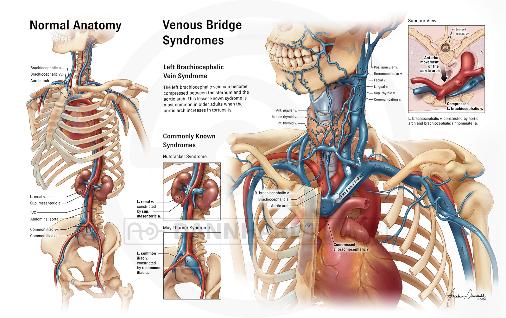
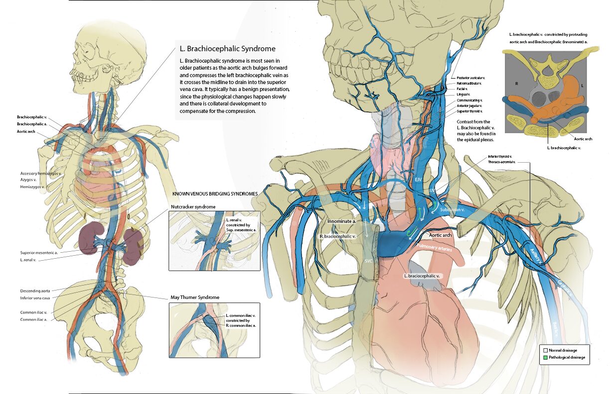
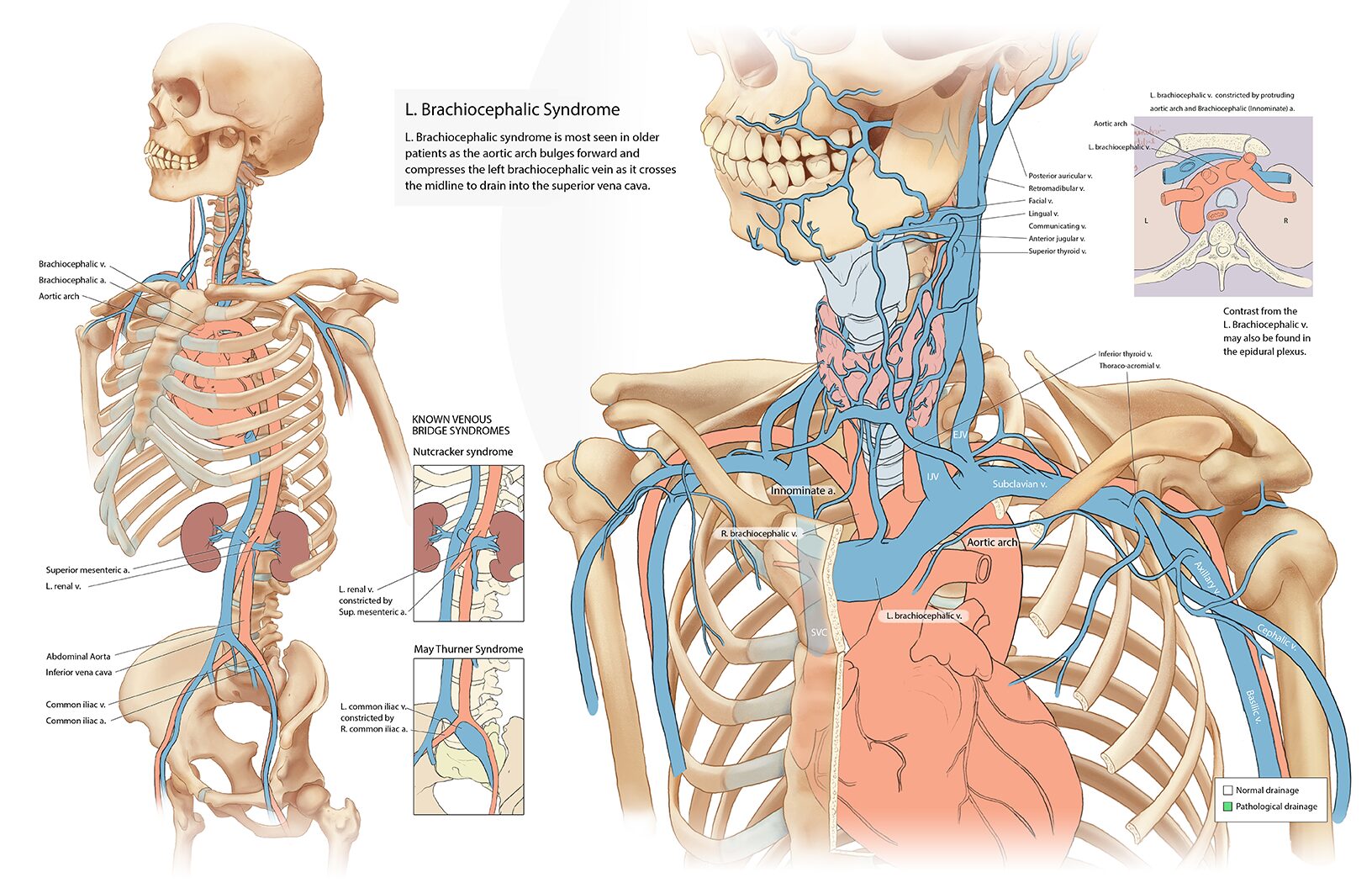
This illustration depicts the venous drainage pathways from the left side of the body as they cross the midline to reach the superior or inferior vena cava. It introduces two known syndromes resulting from the compression of left-to-right venous “bridges”:
-
- Nutcracker Syndrome: Compression of the left renal vein between the aorta and the superior mesenteric artery (SMA), causing renal venous hypertension.
- May-Thurner Syndrome: Compression of the left common iliac vein beneath the right common iliac artery, resulting in leg venous hypertension.
This illustration then describes the rarely described Left Brachiocephalic Syndrome, which results from compression of the left brachiocephalic vein between the aortic arch and sternum, leading to venous congestion.
Research
Research
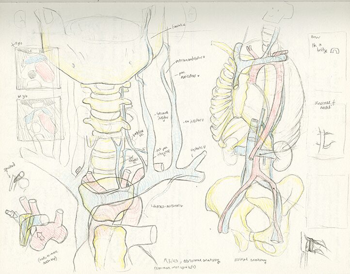
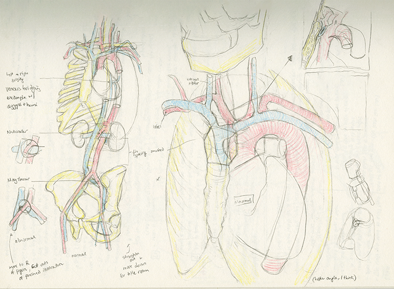
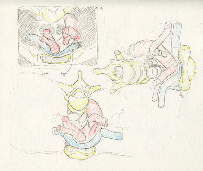
In the drafting phase, I created thumbnails of the relevant anatomy and engaged in discussions about Left Brachiocephalic Syndrome with content expert Dr. Gailloud. He provided valuable insights into the syndrome’s causes, which had not been previously published. Dr. Gailloud also shared several CT screenshots illustrating the disorder. Armed with this information and a solid understanding of normal anatomy, I proceeded to draft multiple layout options for the illustration.
Drafts
Drafting
Once the layout was selected, I created a rough draft followed by a clean draft of the illustration. I then presented it to Dr. Gailloud to ensure that all relevant vessels and anatomical details were accurately depicted, particularly those where significant reflux of contrast would be observed in patients with Left Brachiocephalic Syndrome. Additionally, I sought his confirmation that the accompanying text aligned with the objectives of his paper.

