Intracranial Hemorrhage Compartments
Client
Dr. Haris Sair, Lydia Gregg
Year
2024
Media
Adobe Photoshop
Description
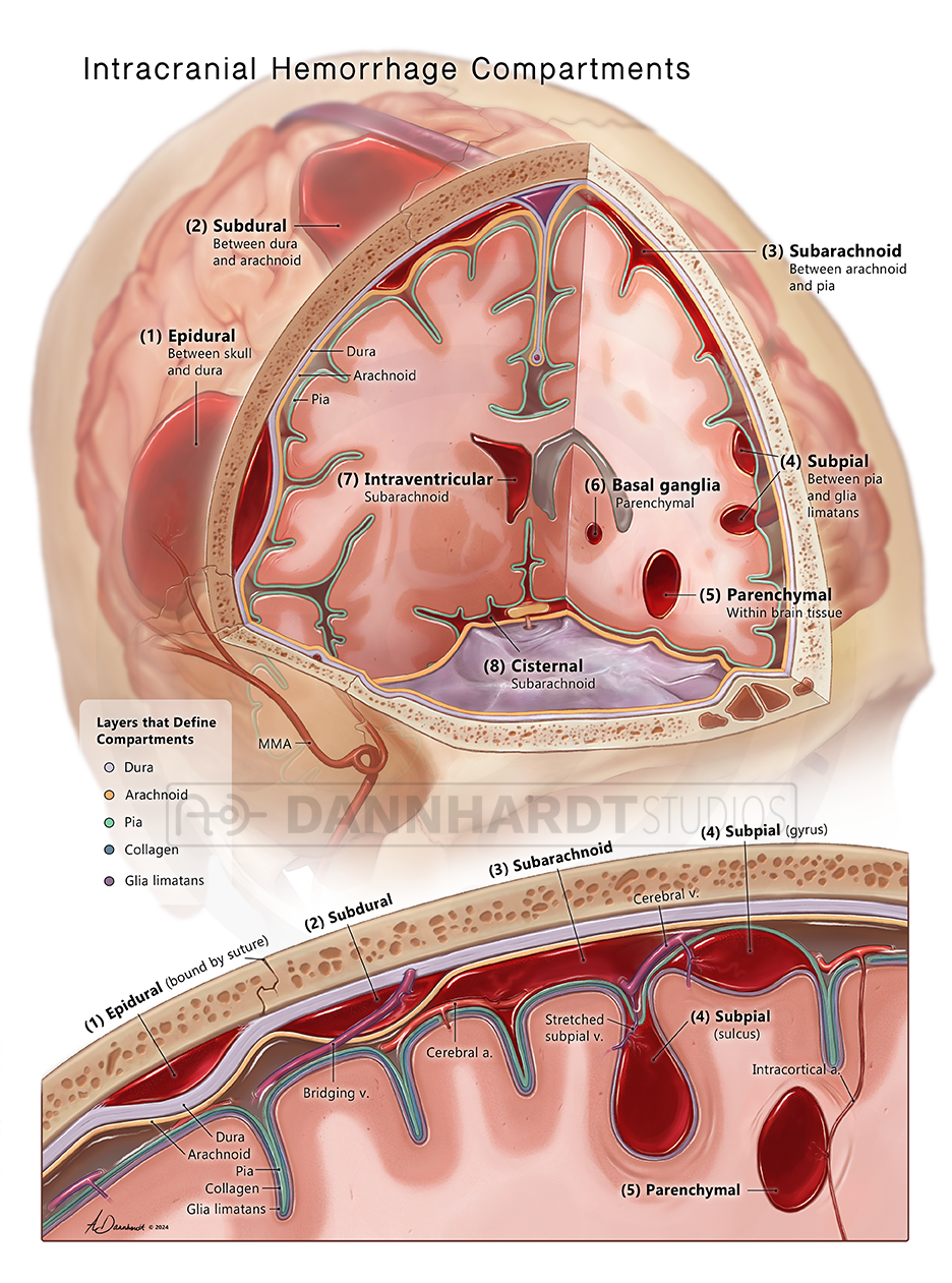
This illustration showcases various hemorrhage types for easy comparison. By cutting the brain past the midline on the sagittal line, the key landmarks of epidural hemorrhage (bound by sutures), subdural hemorrhage (bound by dura), and traumatic lobar parenchymal hemorrhage (commonly around digitate impressions) are highlighted. Having hemorrhages depicted on the brain also enables the highlighting of specific bleeds that, although within a meningeal compartment like the subarachnoid compartment, are identified by distinct names.
Detail of the diagram
Diadetic Diagram
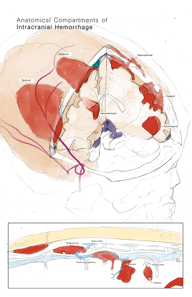
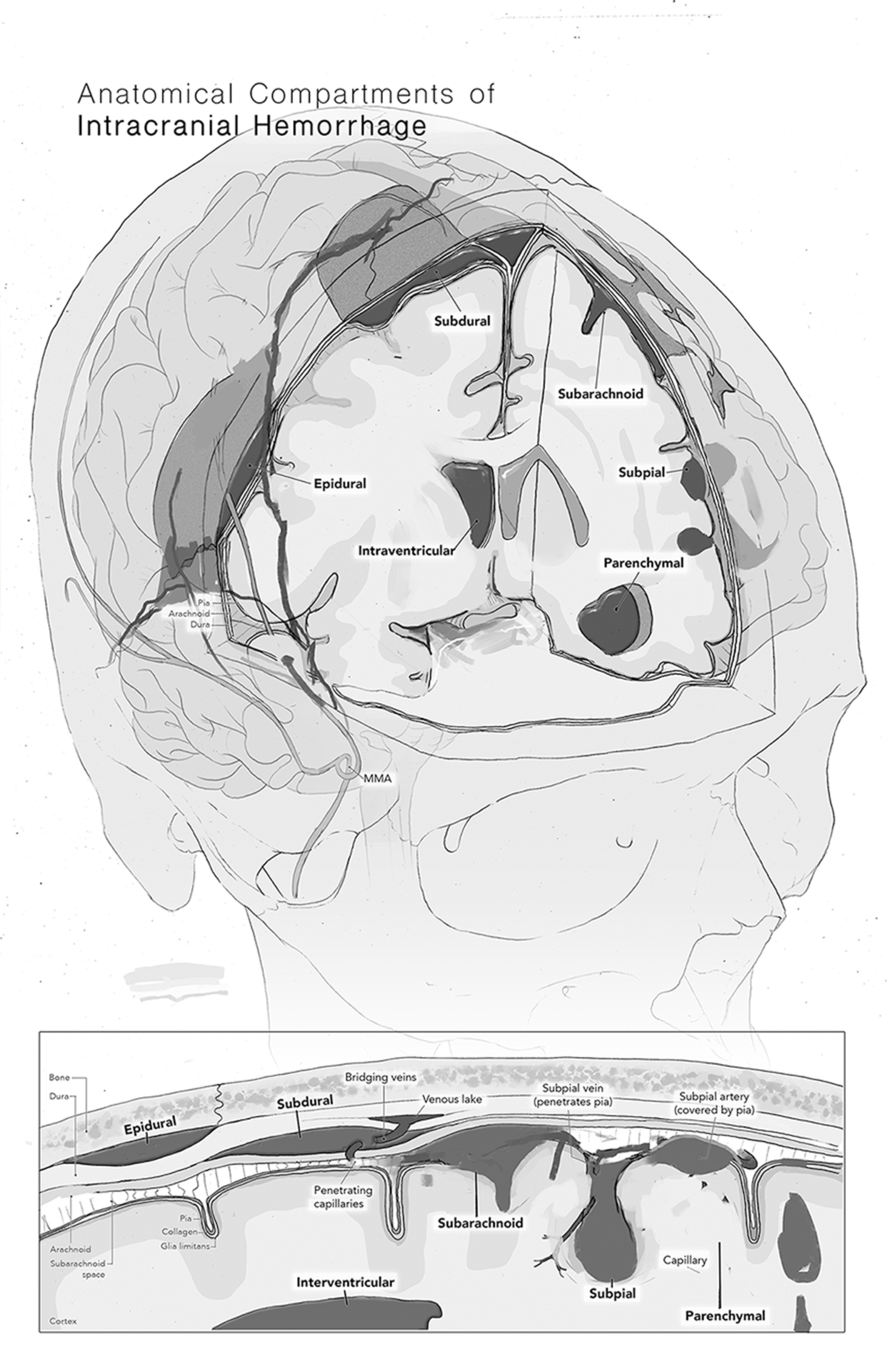
A stylized diagram categorizes hemorrhages based on the five distinct compartments bound by meninges. This simplifies each type and allows inclusion of information about likely sources of the bleed. Notably, this piece includes a visualization of a subpial hemorrhage by illustrating the bleeding into the collagen space between the glia limitans and the pia mater, a rarely described phenomenon.
Drafting
I researched the hemorrhage compartments and their clinical presentation using current literature and publicly available medical imaging. Using Slicer, I created a 3D model of a brain and a skull for visual reference, and then iterated until I felt I had a view that effectively showed all hemorrhages. The piece was then rendered in Photoshop.

Thumbnail

Draft


